The Radiology department at PACHS is committed to providing excellent patient care and maintaining high quality imaging with state-of-the-art equipment.
We offer a wide variety of modalities including:
Our highly trained technicians & radiologists are standing by to serve your lifecare needs.
Radiology department
-
Address
3201 1st St Emmetsburg, IA 50536
-
Hours
Monday - Friday 6:30 am - 6:00 pm
Saturday 7:00 am - 3:30 pm
Emergency Coverage 24/7 -
Fax
712.852.5478
Bone Density
Bone Densitometry
Bone densitometry is a specific test to diagnose and monitor treatment of osteoporosis.
Your Exam
What Will Happen During the Exam?
The patient is asked to lie on their back on the imaging table. During the scan the patient will be asked to lie very still while a camera moves over top of the area being scanned.
How Do I Get Ready for a Bone Density Exam?
Discontinue calcium supplements 24 hours prior to exam.
How Long Does the Exam Take?
This test usually takes between 15 and 30 minutes.
Thyroid Scan
A thyroid uptake measures overall thyroid gland function. A thyroid scan shows the structure, size and location of your thyroid and the function of various portions of your gland. This procedure involves two visits to the Radiology Department.
Your Exam
What Will Happen During the Exam?
For the scan, you will lie on your back on an imaging table with the camera positioned above you. We will take several images of your thyroid. Each image takes five or 10 minutes. We may take additional images to look at a certain part of your gland in detail. The imaging procedure will take about 45 minutes.
The uptake procedure measures the absorption of the radioactive iodine by your thyroid gland.
How Do I Get Ready for a Thyroid Scan?
On the first day, you will be asked to swallow a small amount of radioactive iodine in a capsule. This visit should take about 15 minutes. About 4 to 6 hours later you will return for the first measurement of uptake and the scan, which will take about 15 minutes. The next day, you will return for the remainder of the uptake procedures, which will take 15 to 30 minutes.
How Long Does the Exam Take?
This procedure lasts about 45 minutes.
X-Rays
Diagnostic Radiography
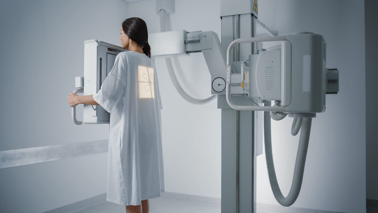
General Radiography is where all X-rays are taken. Our department is equipped with two imaging suites and a portable unit. General radiography is completely film-less. This means that all images are acquired digitally and sent to a workstation to be viewed by the interpreting radiologist.
What Does Film-Less Mean For You?
Several things! First, an exam will be completed much faster than with conventional screen and film radiography. Second, the radiation dose received during an exam will be minimized. Third, storing images digitally allows clinicians within the facility to have fast and easy access to them. Last, we have the ability to transfer your images off-site for emergency interpretation.
Real Time X-Rays
Fluoroscopy
Fluoroscopy is real time X-ray. This means that while X-ray is “on” the image is “live”. Fluoroscopy studies are typically used to evaluate abnormalities in the gastrointestinal (GI) tract such as: Upper GI (UGI), Small Bowel Follow Through (SBFT), and Barium Enemas (BE).
Your Exam
What Will Happen During the Exam?
There are many different types of fluoroscopic studies and each is performed in a different manner. Specifics are given out at the time of scheduling.
How Do I Get Ready for a Fluoroscopic Study?
Most fluoroscopic studies require preparation. Specific instructions will be given at the time of scheduling.
How Long Does the Exam Take?
Again, there are many types of fluoroscopic studies and times vary from 30 minutes to several hours.
CT Scan
Computed Tomography
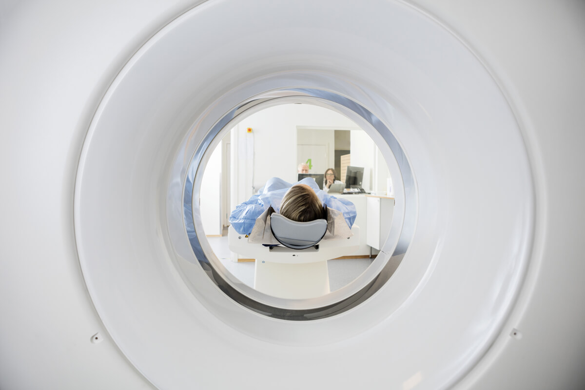
A CT scan is a special X-ray study that takes pictures of the inside of your body. A narrow X-ray beam moves around a body. A picture is quickly taken and a computer interprets the information. The images it produces are “cross-sectional” often referred to as a “slice”, patterned much like slices of bread. A series of these pictures is made to focus on the body part(s) your doctor needs to see.
The Radiology Department at PACHS has a Siemens Somatom Go.All 64 slice scanner. Multi-detector CT has dramatically improved clinicians’ ability to accurately diagnose disease at an early stage. It is a powerful diagnostic tool that uses rotating X-rays to penetrate body tissues, generating multiple slice images, which can detect more than traditional radiography.
In 200 milliseconds, the gantry can rotate around a patients’ entire body – a fast scanning capability that can effectively reduce image distortion of moving parts, such as the heart and lungs. With increased speed, multi-slice CT imaging is especially useful for examining patients who are unable to hold their breath.
With patient care a top priority, Siemens Somatom Go.All uses applications that are specifically designed to consistently deliver the highest image qualities available, while simultaneously achieving significant reductions in radiation and contrast dose.
Your Exam
What Will Happen During the Exam?
Different areas of the body require different scanning techniques. Often the patient is asked to lie on their back and hold his or her breath for short periods. An intravenous injection of contrast material (x-ray dye) may be given.
How Do I Get Ready for a CT Scan?
Preparations may be required and will be explained at the time of scheduling.
How Long Does the Exam Take?
The procedure usually takes between 30 and 60 minutes. Please understand that you may have to wait a few minutes during your scan while the images are being examined. It is important that the pictures contain all necessary information before you are moved from the table.
MRI
Magnetic Resonance Imaging
MRI is an imaging method that uses a strong magnetic field supplied by a magnet and radioactive waves to take pictures of the inside of your body and its chemical make-up. It is a safe imaging method and there are no known biological hazards associated with MRI. An MRI produces pictures that show changes in tendons, ligaments, cartilage and tumors in organs. It is the most sensitive exam to see small tears and injuries to ligaments and muscle. MRI produces high-resolution images that evaluate areas of the body including the brain, spine, bones, and joints.
MRI is provided to our patients though a mobile service (Shared Medical Services) and is available on Sunday and Thursday mornings.
Your Exam
What Will Happen During the Exam?
An MRI requires the patient to lie on a table. The area of the body to be scanned is positioned in the center of the magnet. This determines whether the patient will enter feet first or head first. A small device called a surface coil may be placed over or near the body part being scanned to improve the images.
How Do I Get Ready for an MRI?
MRI scans require clothing without metal and no metal objects such as jewelry, hair accessories, or body piercing.
How Long Does the Exam Take?
An MRI usually takes about an hour.
Nuclear Medicine
Nuclear medicine is an imaging method that obtains pictures by giving the patient a small dose of radioactive material. Images are taken with a special camera that can detect the location of the radioactive material within the body. Nuclear Medicine is used to diagnosis or treat disease, assess tissue function, metabolism or blood flow by injection of radioactive pharmaceutical agents into the patient. These agents attach to organs and body parts, and can be detected by a camera, which records their appearance and function.
Your Exam
What Will Happen During the Exam?
The radioactive material will be given through an injection in the arm or by swallowing a capsule. The area of the body be examined determines how the dose is given. The radiation dose is comparable to a routine x-ray exam and there are no side effects with the radioactive material given. The patient may be asked to lie or sit in front of the camera.
How Do I Get Ready for a Nuclear Medicine Test?
For many of the exams there are no special preparations required, however if preparations are necessary, they will be explained at the time of scheduling.
How Long Does the Exam Take?
Scans range in time from a few minutes to several hours. Some exams require a delay after the material is given, before imaging is started. This is to allow the material to collect at the area of interest.
Pain Management
Our providers from Midwest Radiology and Imaging are skilled in several procedures which can assist in pain management.
These include:
- Epidural Steroid Injection
- Single Nerve Root Injection
- SI Joint Injection
- Arthrograms
- Facet Injections
- Lumbar Spine Myelogram
- Hip Injections
Ultrasound
Ultrasound uses reflected sound waves to acquire images of the organs in the body. An ultrasound and sonogram mean the same thing. An ultrasound does not use x-rays and gives a clear picture of soft tissues that do not show up well on x-ray images.
Ultrasound causes no health problems and may be repeated as often as is necessary if medically indicated.
Ultrasound is the preferred imaging modality for the diagnosis and monitoring of pregnant women and their unborn infants. Also, ultrasound provides real-time imaging, making it a good tool for guiding minimally invasive procedures such as needle guidance and aspiration of fluid elsewhere.
Your Exam
What Will Happen During the Exam?
The patient will be asked to lie down on the exam table and asked to move into different positions during scanning. He or she may also be asked to hold their breath. A warm gel is applied to the part of the body being imaged and an instrument called a transducer is passed over the area by the sonographer.
How Do I Get Ready for an Ultrasound?
Some ultrasound examinations require special preparations. These will be explained at the time of scheduling.
How Long Does the Exam Take?
An ultrasound exam usually takes between 30 and 60 minutes.
Mammograms
3D Mammography
More Accuracy With The Genius™ 3D Mammography.
Why Choose A Genius 3D Mammogram?
A Genius exam detects 41 percent more invasive breast cancers and reduces false positives by up to 40 percent. This means one simple thing: more accuracy.
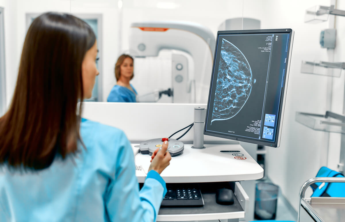
The Genius exam allows doctors to see masses and distortions associated with cancers significantly more clearly than conventional 2D mammography. Instead of viewing all of the complexities of your breast tissue in a flat image, as with conventional 2D mammography, fine details are more visible and no longer hidden by the tissues above or below
Your Exam
What Will Happen During the Exam?
The 3D mammogram is very similar to having a conventional 2D mammogram. The technologist will position you, compress your breast, and take images from different angles.
With 3D mammography, there is no additional compression required, and it only takes a few seconds longer for each view.
The technologist will view the images of your breasts at the computer workstation to ensure quality images have been captured for review. A radiologist will then examine the images and report results to your physician.
What About Radiation?
Very low x-ray energy is used during the exam, just about the same as a film-screen mammogram. The total patient dose is within the FDA safety standards.
How Do I Get Ready for a Mammogram?
You should not use any lotion, powder or deodorant on your breasts or underarms before your mammogram. If you arrive for your appointment wearing any of the above it is okay but, you may be asked to remove it prior to imaging.
How Long Does the Exam Take?
A routine mammogram usually takes between 20 and 30 minutes.
Free Breast & Cervical Cancer Screenings For Women Ages 40-64
Early Detection Saves Lives!
Palo Alto County Community Health System is offering no-cost breast and cervical cancer screenings for women through the Iowa Breast and Cervical Cancer Early Detection Program, which is funded by the Centers for Disease Control and Prevention.
The program includes no-cost screening examinations for breast and cervical cancer and follow-up services, referrals and assistance necessary for medical treatment, for women ages 40- 64 years who meet the program qualifications. In most cases, the screenings can be done using your local physician.
To see if you qualify for this program, please call Candy Bisenius, RN at 712-852-5419.
Peek-A-Boo Imaging
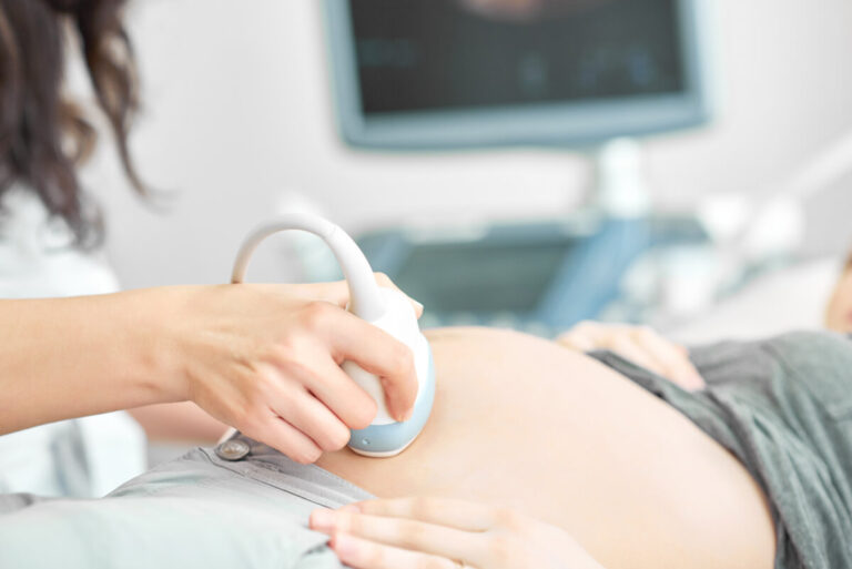
Waiting for the arrival of your new baby is a time of excitement and wonder. Palo Alto County Hospital’s Peek-a-Boo Obstetrical Imaging offers a wonderful opportunity for you and your loved ones to see and bond with your baby before he or she is born.
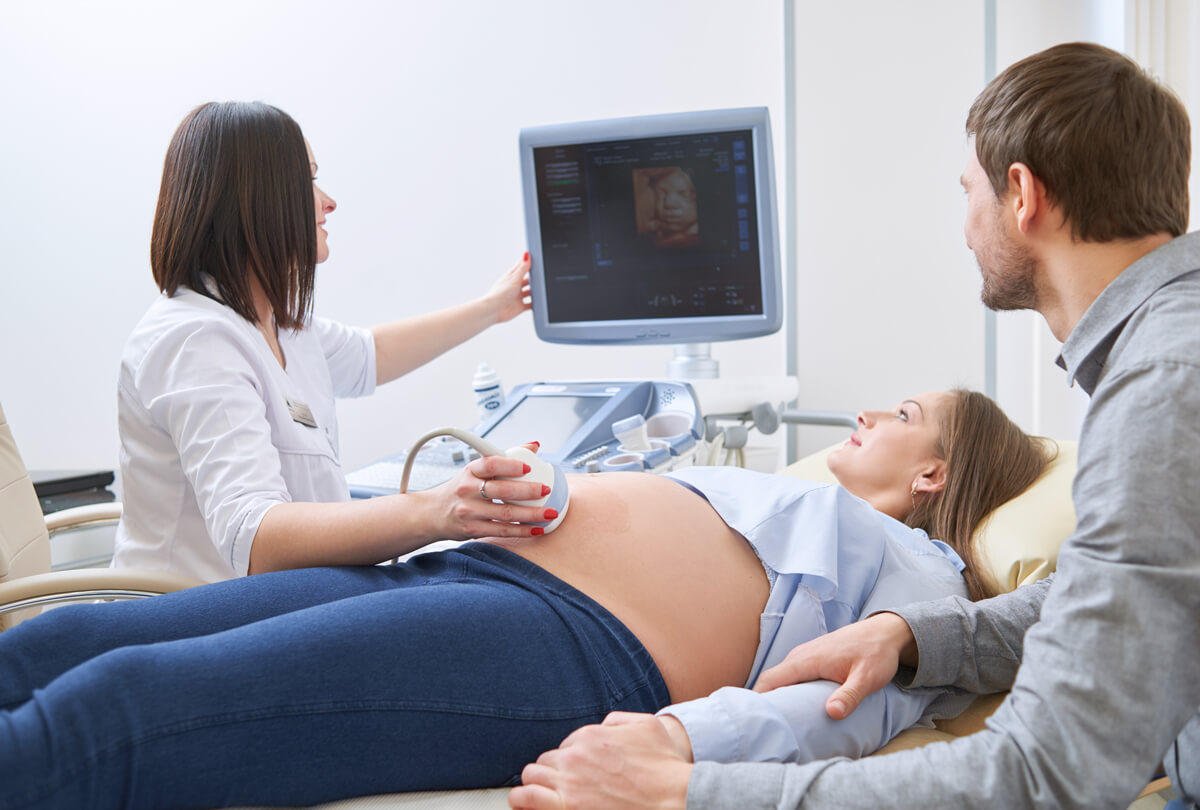
Using state-of-the-art ultrasound imaging equipment, you and your family will watch your baby on a large LCD flat panel in our ultrasound suite. Peek-a-Boo ultrasound sessions are performed by the same sonographer(s) that perform all of our diagnostic ultrasound examinations.


Common Questions
Can I Make This Ultrasound Appointment on My Own, Or Does It Need to be Ordered by My Physician?
The purpose of this ultrasound session is to allow you and your loved ones to see and bond with your baby. This is not a medical diagnostic ultrasound. Therefore, you are welcome to schedule the appointment on your own.
However, all clients must be under the active care of a physician or midwife and the provider must be aware of your appointment.
How Far in Advance Should I Make My Peek-A-Boo Ultrasound Appointment?
We generally have availability within a couple of weeks notice. The appointment may be scheduled in person or by calling our department. Please be sure to specify that you are requesting an appointment for Peek-a-Boo ultrasound.
When Is the Best Time to Come in for My Peek-A-Boo Imaging Appointment?
Babies look and image differently at various stages of pregnancy. The best time for visualization of your baby’s facial features is between 26 and 31 weeks. During this time of pregnancy, your baby has a nice amount of fat and development, which helps to define the face and other features.
Does This Take Place of the Ultrasound Exam(s) Ordered By My Provider?
ABSOLUTELY NOT! Women seeking an elective, non-diagnostic ultrasound must already be receiving treatment with a health care provider for prenatal care. Please note, at no time is this exam to be used in place of a complete diagnostic ultrasound.
Can I Bring Family and Friends to My Ultrasound Session?
Yes! We invite you to bring your loved ones with you to this ultrasound experience. We kindly ask that no food or drinks be brought into the ultrasound suite.
Do I Need a Full Bladder?
No, you do not need a full bladder for this ultrasound session. While we always encourage you to drink water and stay well hydrated, a full bladder is not required for this non-medical ultrasound.
How Much Does the Peek-A-Boo Imaging Session Cost?
The session cost is $50.00.
Payment in full is required in advance by cash or check.
Will This Ultrasound Be Covered By My Insurance?
No, this is an elective service that is not covered by insurance. Your insurance will not be billed.
What If Images Are Unable to Be Obtained?
We cannot guarantee that your pictures will be similar to those you might have seen elsewhere or even from our imaging service. Every baby and mother images differently, depending on gestational age, baby’s position, amount of amniotic fluid, and mother’s condition.
However, if the sonographer determines that the images cannot be obtained in the initial session, one limited follow-up session may be scheduled at no additional fee.
How Do I Make an Appointment for Peek-A-Boo Imaging?
To make an appointment call the Palo Alto County Hospital Radiology Department at 712.852.5416.
How Long Does the Ultrasound Take?
Your appointment is scheduled for 45 minutes. Upon arrival, you will need to fill out two forms. The actual imaging session will take approximately 30 minutes.
